Utilization of the ExVive human kidney tissue model of the proximal tubule to assess nephrotoxicity across a diverse set of chemical classes
Publication Summary:
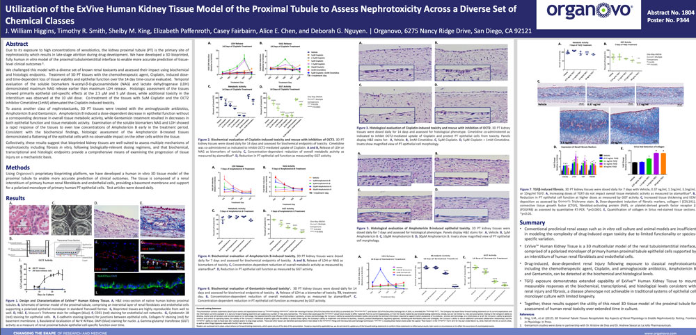
Due to its exposure to high concentrations of xenobiotics, the kidney proximal tubule (PT) is the primary site of nephrotoxicity which results in late-stage attrition during drug development. We have developed a 3D bioprinted, fully human in vitro model of the proximal tubulointerstitial interface to enable more accurate prediction of tissue level clinical outcomes. We challenged this model with a diverse set of known renal toxicants and assessed their impact using biochemical and histologic endpoints. Treatment of 3D PT tissues with the chemotherapeutic agent, Cisplatin, induced dose and time-dependent loss of tissue viability and epithelial function over the 14 day time-course evaluated. Temporal evaluation of the soluble biomarkers N-acetyl-β-D-glucosamindade (NAG) and lactate dehydrogenase (LDH) demonstrated maximum NAG release earlier than maximum LDH release. Histologic assessment of the tissues showed primarily epithelial cell-specific effects at the 2.5 µM and 5 µM doses, while additional toxicity in the interstitium was observed at the 10 uM dose. Co-treatment of the tissues with 5uM Cisplatin and the OCT2 inhibitor Cimetidine (1 mM) attenuated the Cisplatin-induced toxicity. To assess another class of nephrotoxicants, 3D PT tissues were treated with the aminoglycoside antibiotics, Amphotericin B and Gentamicin. Amphotericin B induced a dose-dependent decrease in epithelial function without a corresponding decrease in overall tissue metabolic activity, while Gentamicin treatment resulted in decreases in both epithelial function and tissue metabolic activity. Examination of the soluble biomarkers NAG and LDH showed a rapid response of the tissues to even low concentrations of Amphotericin B early in the treatment period. Consistent with the biochemical findings, histologic assessment of the Amphotericin B-treated tissues demonstrated a flattening of the epithelial cells with no observable impact on the other cells within the tissue. Collectively, these results suggest that bioprinted kidney tissues are well-suited to assess multiple mechanisms of nephrotoxicity including fibrosis in vitro, following biologically-relevant dosing regimens, and that biochemical, transcriptional and histologic endpoints provide a comprehensive means of examining the progression of tissue injury on a mechanistic basis.
View Publication
