Monocrotaline toxicity in 3D-bioprinted human liver tissue
Publication Summary:
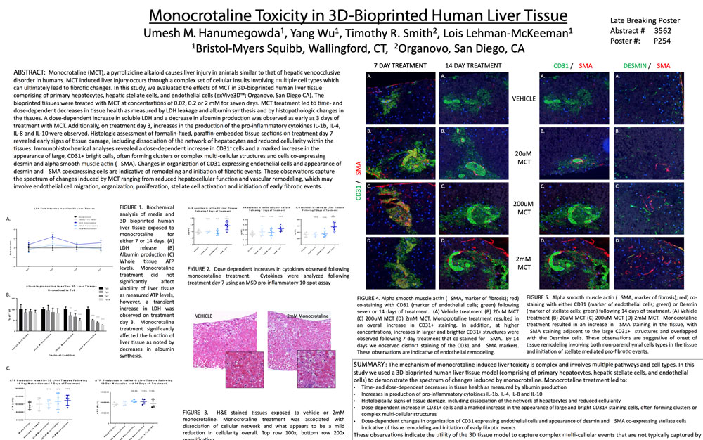
Monocrotaline (MCT), a pyrrolizidine alkaloid causes liver injury in animals similar to that of hepatic venoocclusive disorder in humans. MCT induced liver injury occurs through a complex set of cellular insults involving multiple cell types which can ultimately lead to fibrotic changes. In this study, we evaluated the effects of MCT in 3D-bioprinted human liver tissue comprising of primary hepatocytes, hepatic stellate cells, and endothelial cells (ExVive™; Organovo, San Diego CA). The bioprinted tissues were treated with MCT at concentrations of 0.02, 0.2 or 2 mM for seven days. MCT treatment led to time- and dose-dependent decreases in tissue health as measured by LDH leakage and albumin synthesis and by histopathologic changes in the tissues. A dose-dependent increase in soluble LDH and a decrease in albumin production was observed as early as 3 days of treatment with MCT. Additionally, on treatment day 3, increases in the production of the pro-inflammatory cytokines IL-1b, IL-4, IL-8 and IL-10 were observed. Histologic assessment of formalin-fixed, paraffin-embedded tissue sections on treatment day 7 revealed early signs of tissue damage, including dissociation of the network of hepatocytes and reduced cellularity within the tissues. Immunohistochemical analyses revealed a dose-dependent increase in CD31+ cells and a marked increase in the appearance of large, CD31+ bright cells, often forming clusters or complex multi-cellular structures and cells co-expressing desmin and alpha smooth muscle actin ( SMA). Changes in organization of CD31 expressing endothelial cells and appearance of desmin and SMA coexpressing cells are indicative of remodeling and initiation of fibrotic events. These observations capture the spectrum of changes induced by MCT ranging from reduced hepatocellular function and vascular remodeling, which may involve endothelial cell migration, organization, proliferation, stellate cell activation and initiation of early fibrotic events.
View Publication