Modeling drug-induced hepatic fibrosis in vitro using three-dimensional liver tissue constructs
Publication Summary:
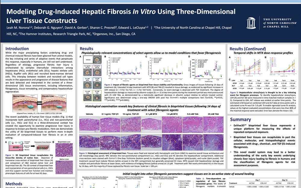
While the major precipitating factors underlying drug- and chemical-induced fibrosis have been gleaned from animal models, the key initiating and series of adaptive events that perpetuate this response, especially in humans, are still not well understood. Regardless of etiology, progressive fibrotic liver injury is orchestrated by complex intercellular interactions among hepatocytes (HCs), endothelial cells (ECs), hepatic stellate cells (HSCs), Kupffer cells (KCs) and recruited bone-marrow derived cells. This interplay between resident and recruited cell types results in the appearance and progression of disease features that are best detected and interpreted in the context of a three-dimensional (3D) tissue environment, including inflammation, fibrogenesis, tissue remodeling, and compensatory hepatocellular regeneration. The recent availability of human liver tissue models that incorporate both parenchymal (i.e., HCs) and non-parenchymal cells (i.e., HSCs and ECs) in a three-dimensional context has created the opportunity to examine progressive liver injury in response to known pro-fibrotic modulators. Here we demonstrate the utility of 3D bioprinted tissues to perform more in-depth evaluation of compound-induced liver fibrosis in an in vitro setting.
View Publication