Inflammatory response of Kupffer cells in 3D bioprinted human liver tissues
Publication Summary:
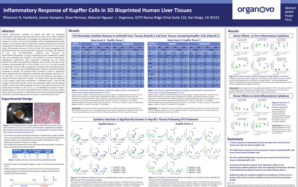
Hepatic inflammation, mediated by Kupffer cells (KC), can exacerbate hepatocellular damage during drug-induced liver injury. KC are often employed in co-culture with hepatocytes to investigate the potential for inflammation in response to stimuli, such as the prototypical inducer lipopolysaccharide (LPS); however, such systems seldom include other nonparenchymal cells and fail to recapitulate the complex 3D interactions present in native liver. In the current study, LPS-mediated activation of KC at 24 and 72 hrs was investigated in 3D bioprinted human liver tissues (ExVive™; Organovo, San Diego, CA). Induction of pro- and anti-inflammatory cytokines was measured via electrochemiluminescence in tissues comprising primary hepatocytes, stellate cells, and endothelial cells (Hep:KC-), and compared to tissues containing KC. Independent experiments were conducted comparing two KC donors (Hep:KC+D1 [male] and Hep:KC+D2 [female]) with all other cell donors held constant. LPS stimulated TNF-α, IL-1β, IL-12p70, IL-10, IL-2, IL-13, and IL-4 levels in Hep:KC+D1 tissues at 24hrs compared to untreated, with sustained induction to 72hrs. TNF-α, IL-10, and IL-8 exhibited greater induction in Hep:KC+D1 tissues compared to Hep:KC-. All cytokines were increased at 24hrs in Hep:KC+D2 tissues treated with LPS compared to untreated, with sustained induction of IL8, IL-1β, IFN-γ, IL-2, and IL-12p70. IL-8, IL-6, and IL-10 induction was greater in Hep:KC+D2 tissues compared to Hep:KC-. To compare donor-specific responses to LPS, cytokine levels were normalized to untreated Hep:KC-, and the fold change in LPS-treated Hep:KC+ tissues was calculated. Patterns of LPS-induced cytokine release were distinct between KC donors, with greater induction in the female donor. However, IL-1β, IL-10, IL-2, IL-13 exhibited no variation in donor specific response. The current data demonstrate the ability to measure a robust donor-specific KC response to inflammatory stimuli, thus enabling investigation of immune-mediated drug-induced liver injury in a 3D human liver tissue.
View Publication