Functional characterization of three-dimensional (3D) human liver tissues generated by an automated bioprinting platform
Publication Summary:
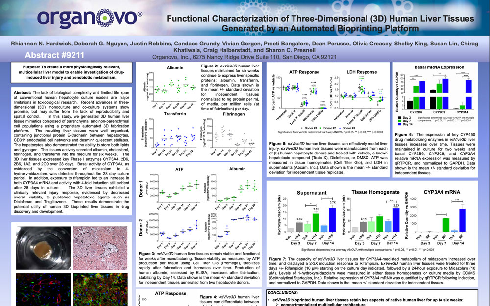
The lack of biological complexity and limited life span of conventional human hepatocyte culture models are major limitations in toxicological research. Recent advances in three-dimensional (3D) monoculture and co-culture systems show promise, but may suffer from the lack of reproducibility and spatial control. In this study, we generated 3D human liver tissue mimetics composed of parenchymal and non-parenchymal cell populations using a proprietary automated 3D fabrication platform. The resulting liver tissues were well organized, containing junctional protein E-Cadherin between hepatocytes, CD31+ endothelial cell networks and desmin+ quiescent stellates. The hepatocytes also demonstrated the ability to store both lipids and glycogen. The tissues actively secreted Albumin, cholesterol, fibrinogen, and transferrin into the medium for six weeks. The 3D liver tissues expressed key Phase I enzymes CYP3A4, 2D6, 2B6, 1A2, and 2C9 over 28 days. Basal activity of CYP3A4, as evidenced by the conversion of midazolam to 4-hydroxymidazolam, was detected throughout the 28 day culture period. In addition, exposure to rifampicin led to an increase in both CYP3A4 mRNA and activity, with 4-fold induction still evident after 28 days in culture. The 3D liver tissues exhibited a clinically relevant injury response, evidenced by decreased overall viability, to published hepatotoxic agents such as Diclofenac and Troglitazone. These results demonstrate the potential utility of human 3D bioprinted liver tissues in drug discovery and development.
View Publication