Development of 3D bioprinted human breast cancer for in vitro screening of therapeutics targeted against cancer progression
Publication Summary:
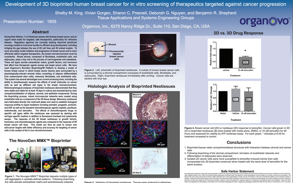
During their lifetime, 1 in 8 American women will develop breast cancer, and an urgent need exists for targeted, safe therapeutics, particularly for refractive disease. Regulatory agencies are currently seeking improved preclinical oncology models to overcome hurdles to efficient drug development, including bridging the gap between the use of 2D cell lines and 3D animal models. To more accurately model initiation and progression of breast cancer and to more efficiently define targeted therapeutics, the breast microenvironment must be considered. Breast stroma, composed of fibroblasts, endothelial cells, and adipocytes, plays a key role in the process of carcinogenesis and metastasis. These cell types secrete extracellular matrix, growth factors, and hormones that affect how therapeutic agents access and target cancer cells. We have used Organovo’s NovoGen Bioprinting® Platform to develop a model of human breast cancer in which breast cancer lesions were surrounded by a physiologically-relevant stromal milieu consisting of adipose (differentiated from mesenchymal stem cells), mammary fibroblasts, and endothelial cells. This system has several advantages over current screening tools, including the ability to simultaneously measure the effects of small molecules on cancer cells as well as different cell types in the breast microenvironment. Histomorphological analyses of bioprinted neotissues demonstrated that they were stable and viable for at least 14 days in culture and characterized by clear compartmentalization of adipose, stromal, and epithelial components. During the bioprinting process, robust microvascular networks were created using endothelial cells as a component of the 3D tissue design. Mammary neotissues were fabricated directly into multi-well plates and used to establish biological response profiles to signal mediators including estradiol, progestin, prolactin, and HGF as well as the standard chemotherapeutic agents cisplatin, paclitaxel, methotrexate, and tamoxifen. The effects of chemotherapeutic drugs on specific cell types within the neotissues was assessed by staining with cell-type specific markers in addition to fluorescent live/dead and cytotoxicity assays. The response of the 3D breast neotissues to growth factors, hormones, and chemotherapeutic agents was compared to the response of 2D breast cancer cell lines. This model can thus be used to screen new anti-cancer targets with better efficiency and accuracy for targeting of cancer cells in the context of the in vivo microenvironment.
View Publication