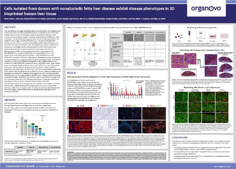
Cells isolated from donors with nonalcoholic fatty liver disease exhibit disease phenotypes in 3D bioprinted human liver tissue
Publication Summary:
The identification of targets and biomarkers and development of therapeutics for nonalcoholic fatty liver disease (NAFLD) may be accelerated by the use of well-characterized primary cell and tissue reagents, as well as improved in vitro human cell-based disease models, including three-dimensional (3D) bioprinted liver tissue. The characteristics of donors from which the cells are isolated, and especially their stage on the NAFLD continuum, are likely to influence the resulting performance in two-dimensional (2D) and 3D models. Evaluation of individual cell type characteristics, and their performance when combined in a tissue coculture model, could enable development of in vitro models more representative of specific patient populations and disease phenotypes.
RNA sequencing (RNA-seq) was performed on (5) non-diseased and (5) NAFLD liver tissues with NAFLD Activity Score (NAS) of 3 or more, revealing clear separation of non-diseased vs. NAFLD tissues and differential expression of fibrosis related genes. Histological analyses performed on tissue microarrays revealed consistent altered distribution patterns of hepatic stellate cells (HSC), with differential activation of HSC. Hepatocytes and non-parenchymal cells (NPC) (HSC, endothelial cells, and Kupffer cells), were isolated from non-diseased donors and from donors with NAS of 3 or more. The isolated cells were characterized with respect to viability, growth kinetics, cytokine production, and phenotype. 3D bioprinted liver tissue was generated using either NPCs isolated from diseased donors combined with non-diseased hepatocytes, or hepatocytes isolated from diseased donors combined with non-diseased NPCs. 3D bioprinted liver tissue generated using NPCs from a diseased donor exhibited accelerated collagen deposition (by trichrome stain) and HSC activation (by α-SMA staining) in comparison to bioprinted liver tissue generated with non-diseased tissue donors. Tissue generated using hepatocytes from a diseased donor exhibited steatosis induction.
Characteristics of the tissue of origin for cells used for in vitro models, including disease status, influence the performance of the cells and the utility of the resulting model. Thus, characterization of cell donors could enable development of in vitro models more representative of specific patient populations and disease phenotypes.
View Publication
