Bioprinted three dimensional human liver contructs provide a model for interrogating liver biology
Publication Summary:
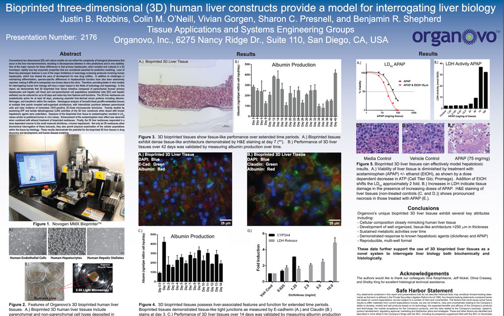
Conventional two dimensional (2D)
cell culture models do not reflect the complexity of biological phenomena that
occur in the liver microenvironment, resulting in discrepancies between in
vitro predictions and in vivo realities. One of the major reasons for these differences
is that primary hepatocytes, when isolated and cultured in a 2D monolayer,
rapidly lose key enzymatic properties that are considered essential for
predictive modeling. Loss of these key phenotypic features is one of the major
limitations in toxicology screening protocols involving human hepatocytes,
which has slowed the pace of development for new drug entities. In addition to
challenges in maintaining differentiation, species-specific differences in
hepatocellular function have also been extensively
reported, making it difficult to extrapolate non-human data to the clinic.
Therefore, providing better in vitro models for interrogating human liver
biology will have a major impact in the fields of toxicology and hepatology. In
this report, we demonstrate that 3D bioprinted liver tissue mimetics composed
of parenchymal (human primary hepatocytes and hepatic cell lines) and
non-parenchymal cell populations (endothelial cells [EC] and hepatic stellates)
can be cultured for up to 42 days and retain key liver features and functions.
The 3D liver neotissues are metabolically active for at least 42 days,
producing essential liver-derived serum proteins including albumin, fibrinogen,
and transferrin within the medium. Histological analysis of formalin-fixed paraffin-embedded
tissues
at multiple time points revealed well-organized architecture, with
intercellular junctions between parenchymal cells and clear evidence of
lumenized, CD31-positive, EC-lined microvascular structures. Toxicity studies
by examining ATP and lactate dehydrogenase (LDH) activities of the 3D liver
constructs when dosed with known hepatotoxic agents were undertaken. Exposure
of the bioprinted liver tissue to acetaminophen resulted in LD50 values similar
to published human in vivo values. Enhancement of the acetaminophen toxic
effect was observed when combined with ethanol treatment of bioprinted
neotissues. Finally, the 3D liver neotissues responded in a dose dependent
manner to the small molecule diclofenac, a known hepatotoxin. Not only do 3D
neotissues allow biochemical interrogation of these toxicants, they also permit
physical examination of the cellular populations within the tissue by
histology. These results demonstrate the potential for the bioprinted 3D liver
tissues in drug discovery and development, and human disease modeling.